If you are citizen of an European Union member nation, you may not use this service unless you are at least 16 years old.
You already know Dokkio is an AI-powered assistant to organize & manage your digital files & messages. Very soon, Dokkio will support Outlook as well as One Drive. Check it out today!
In proton therapy, a proton beam is used. This, unlike regular radiation, can be used by the physicians to control and manage cancer. In doing so, damage to the healthy tissue and vital organs is significantly reduced because proton therapy works on the principle of selective cell destruction. A proton beam has a low scatter effect in the tissue, and stays focused on the tumor.
Proton Therapy is a type of therapy that uses a beam of protons to irradiate diseased tissue, usually cancer. The major advantage of proton therapy is being able to precisely localize the radiation dosage when compared with other types of external beam radiotherapy.
Questions: Ashley Mizuno: -How does it work?
In photon therapy, the energy of the protons can be directed and delivered in the tissues designated by the physicians. This is done by using a 3 dimensional pattern from each proton beam that is being used. In doing so, there is greater control and accuracy. The healthy tissues surrounding the cancer site have a less chance of being damaged due to ionization. Because of this directed therapy, a wide range of cancers can be treated. These include tumors in the head and neck area, lung, prostate, bladder, spinal cord, gastro malignancies and ocular tumors. Damage to lung and surrounding breast tissues can be avoiding when treating breast cancer with proton therapy.
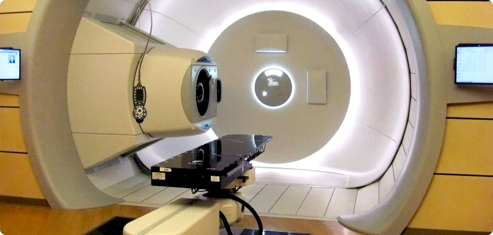
To begin, hydrogen atoms are separated into electrons and protons in a cyclotron. The protons are then sent through a vacuum tube with a pre-accelerator. From here, they make their path to a synchrotron, where the protons are accelerated. A radiofrequency signal delivers a boost of energy here, which then increases the energy of the protons. This energy, in volts, depends on how far it needs to travel in the body. After leaving the synchrotron, the protons move through a transport system that is directed by magnets to the tumor. Each treatment of protons uses a guidance system to steer the beam to the patient. This system monitors the proton beam until it enters the patient. It also conforms to the size/shape of the tumor, a plan that is designed by physician. The system that delivers the proton beam, called the nozzle, is the last path traveled though before entering the patient. This shapes, and spreads out the beam in three dimensions. The location, shape of tumor, depth and tissue density is all taken into consideration when calculating the number and velocity of protons that are delivered. Although all this is occurring, patients only see a revolving cone-shaped device.
In order to be so precise, protons are energized to certain velocities and this determines how deep the proton’s maximum energy will be deposited into the body. Protons slow down as they travel through the body, which increases the protons interactions with orbiting electrons. As protons approach the desired cancer site, there is a maximum interaction with the electrons. Therefore, the maximum energy is released at the designated site. As a result of this process, the healthy tissues surrounding this site receive less damage and the patient can receive a higher, more effective dose, with low side effects. This can improve the patient’s quality of life and provide long term health.
-What the difference is between proton and other types of radiation?
Selective cell destruction is the goal of all types of radiation, but go about it in different ways. Standard x-ray therapy uses photons instead of the heavy, positively charged particles (protons) in proton therapy. It uses x-ray beams in which the amount delivered is designed to provide enough ionization in order to damage all the cancerous cells. Much of this energy from the x-ray beam is being absorbed by the normal tissues near the body’s surfaces as well as beyond the desired cancer site. This is due to the fact that x-rays lack charge and mass. This can lead to unwanted damage to healthy tissues. Another radiation therapy, called fast neutron therapy ,uses high energy neutrons, which can be very effective in killing tumors, but can be equally lethal to normal tissue. Neutrons, pions and heavy ions deposit more energy along their path, consequently causing more damage to the cells they hit.
-What is the process that someone has to go through and the side effects of it?
Before a patient undergoes proton therapy, the patient has to go see a radiation oncologist to see if the patient is a candidate for the procedure. To be a candidate for proton therapy there are some conditions. There is a list of tumors and cancers that proton therapy can work and also there is a weight limitation because of the equipment, people over 360 pounds can not be a good candidate. To find out more if a person is a good candidate for proton therapy it is best to contact the hospital that the person wish to do the therapy and they will have a consultation and be able to receive more information. If the patient is a candidate, the patient will be fitted for an immobilization device where depending on where the location of the tumor the device will be fitted and used for each therapy session. For example, if the location of the tumor is under the neck will have a full body mold that is made up of foam liners which is surrounded by rigid plastic shells. If the location is in the head area, then the patient will be fitted with a custom made mask. There are different types of immobilization devices such as the custom made mask, bite molds, body casts, arm or leg rests.
Once the device is made then the patient has to undergo either a magnetic resonance imaging (MRI) or Computed tomography (CT). Both will scan for the tumor and will create a 3D image of the tumor with its boundaries surrounded by the normal structures. While the patient is undergoing the MRI or the CT they are also wearing the device for treatment planning. A physicists or dosimetrists will create a treatment plan on the computer that will either outline a single or multiple proton beams that will enter at different angles. And with this they can calculate how much radiation dose the tumor will receive.
When the actual therapy starts, the average procedure treatment usually lasts five to seven weeks, depending on the size, type and what stage the tumor is on. The delivery of the proton beam to the patient lasts about one minute but the patient will probably spend around 15 to 20 minutes in the room with the preparations, positioning, and adjustments.
Video of the Process of Proton Therapy:
This therapy is performed on an outpatient basis and the delivery of the proton beam to the patient lasts only about a minute, during this time the patient will not feel any pain or discomfort from the beam. Proton therapy, compared to standard radiation therapy with x-rays, have less frequent and usually less intense side effects because the therapy injures less healthy tissue. Other factors depend on how big of a dose the patient received and if the patient is also receiving chemotherapy at the same time. Common side effects that a patient will encounter are temporary hair loss and skin reactions where the proton beams, fatigue, nausea, loss of appetite, vomiting, or diarrhea.
Kathleen Yip: -What diseases are treated with this kind of therapy?
Proton therapy is focused mainly on cancers with localized tumors (benign and malignant) since a beam is being utilized. My colleagues have previously addressed this point. I have constructed a simple list of the majority of diseases treated with proton therapy:
Body Part Location
Disease
Summary
Other Treatments
Brain and Spinal Cord
AVMs = Arteriovenous Malformations
AVMs are actually defects of the circulatory system. They occur during development sometimes as early as a fetus or right after birth. You can think of them as tangled arteries and veins. They do not allow nutrients to pass from the blood to the tissues in the affected area. They usually don't cause problems until after ages 30-40 years.
Surgery, Embolization (plugging of the AVM)
Isolated Brain Metastases
Metastasis is defined as the spreading of a disease such as cancer. Isolated Brain Metastases are tumors that traveled from somewhere else in the body to the brain.
Surgery followed by chemotherapy
Pituitary Adenomas
Tumors found in the pituitary gland. These tumors can secrete humors or press against the optic nerve distorting a patient's vision.
Microsurgery
Base of Skull
Acoustic Neuromas
They are benign tumors and cause swelling of the 8th cranial nerve which is crucial to our hearing. Sadly it most cases any kind of treatment is unlikely to save useful hearing abilities.
Removal of 8th cranial nerve
Chordomas
A rare form of bone cancer that originates in the lower spinal column or tip of the spine. The location of the chordomas make treatment options limited. They are usually close to the brain stem or central nervous system tissue.
Surgery and chemotherapy
Chondrosarcomas
A type of rare cancer arising from cartilage. Found mostly in adult males. Like the Chordomas, Chondrosarcomas are very close to vital tissues (brain stem and central nervous system).
Surgery
Meningiomas
Tumors of the central nervous system, originating from the arachnoid cells of the arachnoid villi in the meninges. Meninges surround the brain and spinal cord. They are usually benign but can be malignant.
Surgery
Eye
Uveal Melanomas
Also called Choroidal Melanomas. These are malignant tumors found in the eyes. The tumors develop in the pigment cells (melanocytes) of the eye.
Enucleation = Removal of the entire eye.
Head and Neck
Nasopharyngeal Carcinoma
This cancer orginates in the nasopharynx. That is where the nasal passages and auditory tubes meet the remainder of the upper respiratory tract. Common in areas with high incident rates of viral, dietary and genetic problems.
Chemotherapy
Oropharynx Cancer
This is when tumors form in the oropharynx. The oropharynx is located in the middle of the pharynx or throat. It encompases the back 1/3 of the tongue, the soft palate, the side and back walls of the throat and the tonsils.
Surgery
Chest and Abdomen
Stage 1 Lung Cancer
This is in the early stages of Lung Cancer. Usually surgery is utilized at this stage but for patients with decreased health (severe heart or lung disease) proton therapy is the way to go.
Surgery
Pelvis
Prostate Cancer
This cancer has tumors forming on the prostate (small round gland which produces seminal fluid, fluid that transports sperm). The prostate gland is very close to a man's urethra. One of the most common cancers for American men. Some proton therapy facilities dedicate the majority of their treatment appointments to prostate treatment since it is so prevalent.
Surgery
Pediatrics
Brain Tumors
Brain tumors can be either malignant or benign. There are the most common solid tumors found in children. Proton therapy is used to minimize the damage to the developing tissue in children.
Surgery, chemotherapy, steroids or anti-seizure medication (swelling)
Orbital and Ocular Tumors
There are many vital organs and parts within the orbit of our body. Precision in treating diseases within this region is key! If even the smallest part is damaged this can lead to complete loss of the eye.
Enucleation = removal of the entire eye (for ocular tumors), Cryotherapy (freezing), Chemotherapy
Sarcomas of the Base of Skull and Spine
Include Chordomas and Chondrosarcomas like I talked about before. The location of diseases makes treating them very difficult and again precise targeting is needed.
Chemotherapy
-Cost (to build and cost of treatment) Proton therapy maybe be one of the most expensive therapies available today. When the University of Pennsylvania was constructing it's brand new proton center the price tag was $144 million! It is as long as a football field with five treatment rooms. It took around 2-3 years to complete. The University of Florida has a 98,000 square foot Proton Therapy facility costing $125 million. In June of this year Scripps Health Center based out in San Diego set plans in motion to build a $185 million proton therapy facility in Mira Mesa, CA. They hope to be open for business by the spring of 2013. The equipment needed for this gigantic (102,00 square foot) facility will top out at $90 million. The cost of conventional x-ray radiation therapy machines is only $3 million each. Most hospitals and clinics have several x-ray machines. The cost for the combined x-ray machines still doesn't even come close to the amount for a proton therapy facility.
According to an article I found in Forbes Magazine Medicare pays twice as much for a complete treatment of protons as for x-rays: $34,000 for eight weeks of therapy versus $16,000. Numbers from Forbes article: -$841 is the cost to Medicare for one session of proton therapy -$411 is the cost to Medicare for one session of conventional x-ray therapy It was found that private insurance companies also shell out some dough to cover proton treatments. However they usually require pre-approval. Even though proton therapy usually costs more than conventional radiation therapy, it still costs less than surgery.
Proton therapy is based on about fifty years extensive and positive experience of the effect of proton beams on diseased and healthy cells in the body. To date, more than 50,000 patients have been treated with protons at about 30 centers worldwide, predominantly those with eye tumors, brain tumors, or tumors in the area of the head and neck and pelvis. This broad clinical experience has shown very clearly that, compared to photon therapy, it is the spatial precision in applying the radiation, which is of decisive importance. This provided the motivation for PSI specialists to develop a new proton therapy facility for high-precision irradiation of deep-seated tumors. The PSI gantry has been in operation since 1996 and by middle of 2008, more than 350 patients had been treated.
Melanomas of the eye In 1984 the first treatments with protons of eye tumors in Western Europe were carried out at PSI. By end of August 2008 we had treated more than 5000 malignant eye tumors with the OPTIS proton therapy facility. The therapy success rate includes more than 98% of the tumors being cured and in 90% of the cases the eye could be saved.
Spot-Scanning for deep-seated tumors The success rate of the eye treatments and the good experience with proton therapy at the Harvard Cyclotron Laboratory in Boston provided the motivation for the PSI specialists to develop a new proton therapy facility for high precision radiation of deep-seated tumors. For this installation a new treatment technique has been developed, where a pencil beam of protons is computer controlled in such a way that high dose spots of protons can be positioned very precisely, for an exactly specified period of time and at any desired location within the tumor. By superimposing many different spots - about 10,000 within a volume of one liter - the radiation dose can be distributed uniformly over the entire tumor. This is the principle of the Spot –Scanning Technique implemented into the PSI-Gantry.
Meningiomas More than twenty-five patients were suffering from meningiomas. These tumors arise from the coverings of the brain (the meninges) which are located between the bones of the skull and the surface of the brain. They grow slowly, displacing adjacent structures, and may infiltrate the surrounding cranial bone. Depending on location and size, they may cause severe headaches, epileptic attacks, disorders of brain function, episodes of loss of consciousness, paralyses and other neurological defects. If surgical removal is impossible, the precise spatial adjustment of the radiation dose involved in proton therapy offers particular benefits.
Brain tumors About 30 treatments have been carried out for patients with brain tumors (for example: gliomas, ependymome, medulloblastome). Most such tumors arise from glial cells, the supporting cells of the brain tissue, and the more closely they resemble the cells from which they originate, the more likely they are to respond well to treatment. In these cases we refer to tumors as grade 1 and 2. When the tumor cells have developed differently to the original cells, so called (entdifferenziert) then proton therapy cannot promise more than photon radiation as the whole brain must be evenly treated. These grade 3 and 4 tumors and glioblastomas are not part of the treatment program at PSI. These tumors are usually treated in clinics with a combination of chemo- and radiation therapy.
Chondrosarcomas and Chordomas About 200 patients were referred for proton therapy because of chondrosarcomas or chordomas involving the base of the skull or the spinal axis. Such tumors are of connective tissue origin, grow slowly and very seldom metastasis, but destroy the most susceptible adjacent structures, often those which are essential for life. The cranial nerves are located at the base of the skull, the optic nerves are close by, as also is the optic chiasm. Permanent damage to these structures is likely to cause blindness. The brainstem, as a part of the central nervous -system, cannot tolerate persistent pressure from a tumor, far less actual invasion, but it also has limited tolerance towards irradiation. Because the proton beams stop at a calculable depth, depositing most of their radiation load within this stopping region (the Bragg peak), chordomas and chondrosarcomas of the skull base have for many years been treated by irradiation. This has become the subject of a well organized clinical trial conducted mainly with the Harvard cyclotron at Massachusetts General Hospital in Boston. Convincing success has been achieved: for chondrosarcomas local tumor control has been raised from about 40% to over 80%, while for chordomas it is now about 65%. These tumors, like melanomas of the eye, have therefore become an indisputable indication for proton therapy. We have achieved successful tumor control for our patients, together with general well-being and a highly satisfactory quality of life.
Sarcomas Sarcomas – malignant tumors arising from the connective and supporting tissues – also occur in other parts of the human body, for example, in the coccyx, in the lumbar vertebral column, on or within bones and joints and also in muscle tissue. Up until middle of 2008 we treated more than 50 patients, with sarcomas not in the spinal or scull base area. Some of these treatments were in partial combination with surgery and or chemotherapy and appear to be successful so far.
Prostate Cancer Up until the end of 2003, we treated 13 cases of prostate cancers with protons. These treatments were likewise successful, and the patients are doing well. In the years 2004 to 2007 there was not the capacity to treat any prostate cancer patients.
ORL tumors Tumors in the ear, nose and throat region (ORL tumors) often grow in close proximity to the sensitive structures of the brain, the base of the skull or the spinal column. Up to middle of 2008, we had treated more than 20 ORL tumors (including two skin tumors), in partial combination with surgery, chemotherapy and, in some cases, also with conventional radiation.
Children and youth Until middle of 2008 we treated more than 90 children and youth between the ages of one and twenty-years old. About 50 of these between the age of 1 and 5 had to be treated under anesthesia, in order that they remained absolutely still during the treatment. The tumors were located in the scull or the base of the scull and between 14.0 and 74.0 CGE was applied. In some cases this was also in combination with conventional photon radiation treatment.
Age distribution of patients More than 40 % of patients were under 40 years of age, while more than 25% were children
and young people under 20. Such patients derive particular benefit from proton therapy, because the precise delivery of the highest radiation dose within the tumor minimizes damage to the vulnerable and still growing body of the child or young person. It also means that subsequent, secondary tumors are less likely to arise than after photon therapy.
Intensity-modulated proton therapy (IMPT) PSI operates world's only facility able to treat patients with intensity-modulated radiation therapy with protons (IMPT). First patient treated with IMPT was a young male patient, suffering from a chondrosarcoma of the upper thoracic spine in 1999. Once again, our objective was to control the tumor, while giving the best possible protection to the immediately adjacent spinal cord, so as to preserve the working capacity and quality of life of this 36 year-old man. Seven years after the conclusion of the therapy, he is free of symptoms and working full time. In the last three years we have further developed this technique, and in the past years more than 50 patients were treated with IMPT.
Prospects for curing tumors with proton therapy In over 80% of the cases we have treated we have been able to stop the growth and kill the tumor, curing the patient. The remainder of the patients had such an advanced tumor growth or it was metastasized so that despite the tumor being controlled the patient was not cured. However in many cases an alleviation of suffering was achieved, and treatment also slowed advancement of the disease. This was achieved with nearly no side effects. Unfortunately, in some cases the tumors grew again.
Clinical experience – future developments The clinical results achieved so far seem to reliably indicate that the developments in proton radiation techniques can offer patients with certain cancer indications considerable benefits in therapy and healing. Under the project PROSCANthe facility is being expanded and by the end of 2009 we will be able to treat four to five times as many patients with deep seated tumors on a year-round basis. Due to the technical capabilities of the new Gantry 2, moving tumors will also be able to be treated with the new treatment technique.
-What is the length of average treatments?
The procedure is performed on an outpatient basis. For most tumor sites, the average course of treatment is usually five to seven weeks, but rarely, certain tumors treatment may last only a few days. The length of each treatment will vary depending upon the tumor type and stage. The delivery of the proton beam to the patient lasts only about a minute, although the total time spent in the treatment room will be longer (about 15-20 minutes) for positioning and adjustments to the equipment settings.
For daily treatments, the patient enters the treatment room and is fitted with his or her personal immobilization device. The patient is positioned with the aid of laser sights to within a half-centimeter accuracy. The radiation therapist then takes several low-energy diagnostic radiographs (x-rays) or digital images to insure proper alignment. This process is repeated before each treatment. In some cases a fan beam CT system will be used to image the target before each treatment.
Special apertures and filters that are made for each patient are loaded into the beam line. A computer may be used to scan and verify the individual bar codes on these devices. Once positioning and treatment parameters are verified, the radiation oncologist and technologists step out into a control room located next to the treatment room and begin the treatment. After the prescribed radiation dose has been delivered, the computer shuts off the proton beam and the technologists re-enter the room to assist the patient in removing the mask or immobilization device.
-History
The first suggestion that energetic protons could be an effective treatment method was made by Robert R. Wilson in a paper published in 1946 while he was involved in the design of the Harvard Cyclotron Laboratory (HCL). The first treatments were performed at particle accelerators built for physics research, notably Berkeley Radiation Laboratory in 1954 and at Uppsala in Sweden in 1957. In 1961, a collaboration began between HCL and the Massachusetts General Hospital (MGH) to pursue proton therapy. Over the next 41 years, this program refined and expanded these techniques while treating 9,116 patients, before the Cyclotron was shut down in 2002. The first proton therapy center in Western Europe has been in operation at the Paul Scherrer Institute (PSI) in Villigen, Switzerland, since 1984. Following this pioneering work, the Loma Linda University Medical Center (LLUMC) in Loma Linda, California was built in 1990.Later, The Northeast Proton Therapy Center at Massachusetts General Hospital was brought online, and the HCL treatment program was transferred to it during 2001 and 2002.
This is your Sidebar, which you can edit like any other page in your workspace.
This Sidebar appears everywhere on your workspace. Add to it whatever you like -- a navigation section, a link to your favorite web sites, or anything else.


Comments (0)
You don't have permission to comment on this page.